Traditional workflow
4–12 hours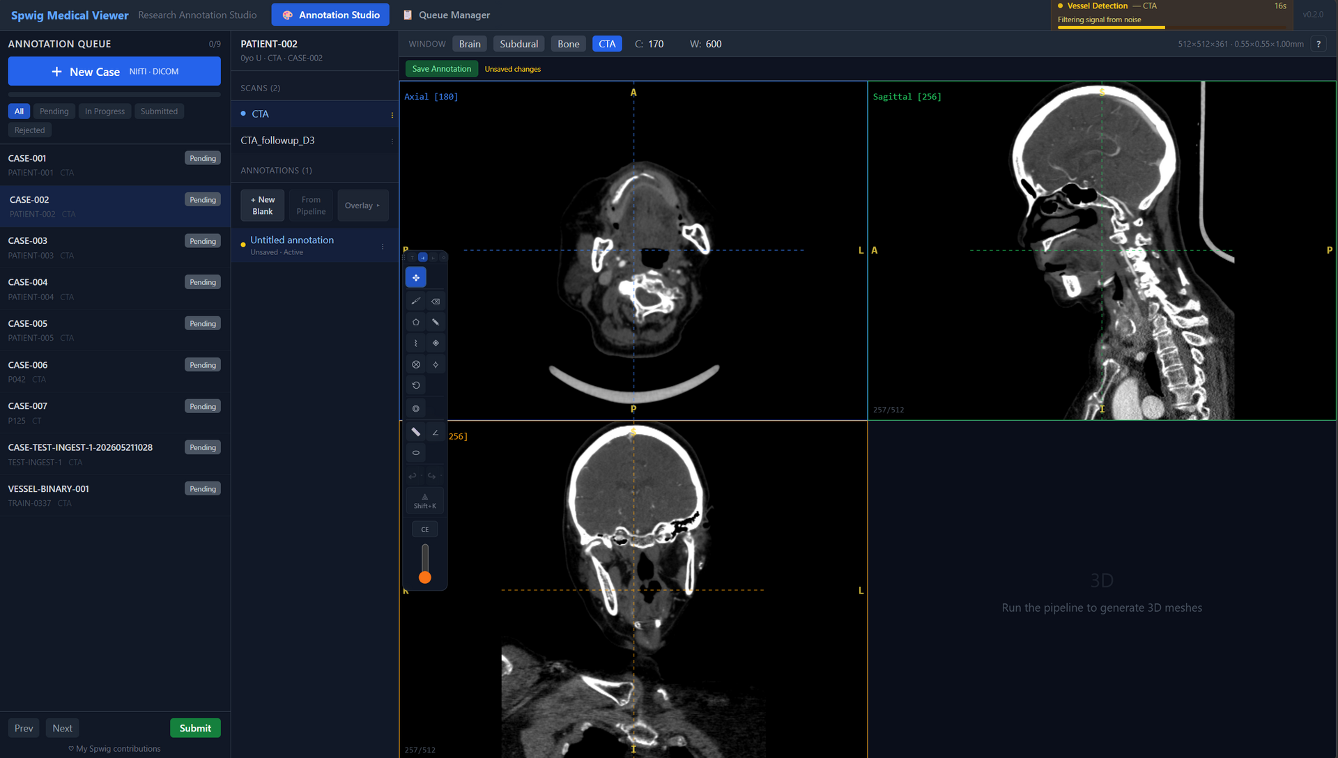
Manual tracing in slice-by-slice 2D, then painful 3D verification. A trained annotator marks every vessel by hand across hundreds of slices.
Open cerebrovascular research tool
Our physics-based pre-annotation engine detects ~90% of cerebrovascular structures automatically. You spend your time correcting edge cases, not tracing arteries from scratch.
Research use only. Not a medical device.

Pre-annotated cerebrovascular structures, ready for review in the Annotation Studio.
The speed gain
Traditional vessel annotation is one of the most time-expensive tasks in cerebrovascular research. Spwig Medical Viewer collapses that into a guided review.
Traditional workflow
4–12 hoursManual tracing in slice-by-slice 2D, then painful 3D verification. A trained annotator marks every vessel by hand across hundreds of slices.
With Spwig Medical Viewer
10–20 minutes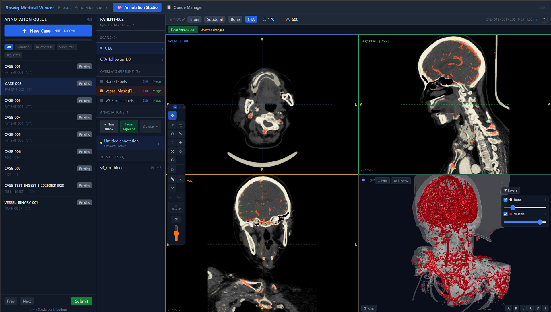
The physics engine pre-annotates ~90% of vessels in under a minute. You correct edge cases with keyboard-driven tools and immediate visual feedback.
Times measured on a 512×512×400 CTA volume, RTX 4090, single annotator.
Features
A GPU ray-cast pipeline detects ~90% of cerebrovascular structures automatically — carotids, vertebrals, basilar, Circle of Willis, MCA/ACA/PCA branches — with scan-adaptive HU calibration. No training data required, no model drift.
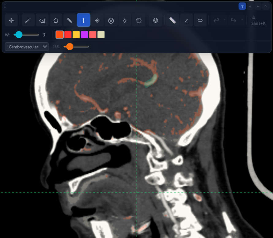
Every tool maps to a keyboard shortcut. Brush, lasso, polygon, Bezier, magic wand — switch tools as fast as you can think. No more hunting for buttons.
Every stroke renders in real time across all four quadview panels and the 3D scene. See exactly what's selected, where it lives in space, and how your edits affect downstream structures.
Annotate directly on the 3D vessel mesh, not just on 2D slices. Click on the basilar artery in 3D, reclassify it, and watch the segmentation update everywhere instantly.
The annotation toolbar
Brush, polygon, lasso, Bezier, magic wand, ROI — every annotation tool is bound to a single key. Switch tools in muscle memory, not by hunting through menus.
Pipeline
GPU ray-cast over the volume identifies all dense structures — bone and contrast-enhanced vessels — in a single sweep.
Hessian geometry classifier separates tubular vessels from sheet-like bone, refined by scan-adaptive HU thresholds.
Active contour gap filler, junction connector, and elliptical cross-section profiler clean up the vessel network.
Open the result in the 3D annotation studio. Correct, label, and export to NIfTI / GLB / JSON.
Install
Your own workstation or GPU laptop
One-click installer for Windows and Linux. Bundles the viewer; downloads CUDA libraries on first launch. Requires an NVIDIA GPU.
Shared lab GPU server
Docker image with one-line bootstrap. Users connect via browser — no client install. Best for labs with central compute.
Research
If Spwig Medical Viewer contributes to a publication, please cite it. A formal preprint with methods detail is in preparation.
Spwig Medical Viewer (Version X.Y.Z) [Software]. Spwig Pte. Ltd. https://medical.spwig.comFree for academic research and education. Sign up for a download link and we'll keep you posted on new releases.